X-ray compatible microfluidics
Microfluidic cells with ultrathin silicon nitride windows for following chemical reactions in liquids, from their first milliseconds, with tender and hard X-rays.
The problem
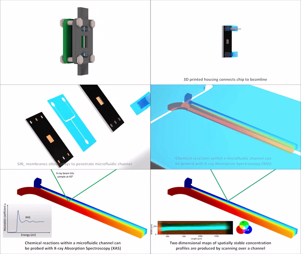
X-ray techniques such as absorption spectroscopy, fluorescence mapping and diffraction reveal the chemical state, structure and distribution of elements in solution, but many important elements (from sodium to iron) have absorption edges in the tender X-ray range, 1–5 keV. These X-rays are strongly absorbed by air, glass and polymer films, so experiments must run in vacuum or helium, and conventional Kapton or glass flow cells attenuate too much of the beam. Probing two solutions at a well-defined time just after they meet is harder still.
How it works
Our devices sandwich 90 µm-deep PDMS microchannels between a 120 nm silicon nitride window and a rigid substrate. The thin window transmits tender and hard X-rays into and out of the channel, so spectra can be recorded either from the fluorescence emitted by the sample or from the beam transmitted through it, and the assembly withstands vacuum. Laminar-flow mixing geometries (Y-junction and flow focusing) create stable diffusion interfaces, so distance along the channel corresponds directly to reaction time; droplet generators and silicon-backed variants extend the range of experiments.
Performance
Suitable for tender (1–5 keV) and hard X-rays, in vacuum
Measurements in both fluorescence and transmission geometries
Compatible with a range of X-ray techniques, including absorption spectroscopy, fluorescence mapping and diffraction
Stable operation under continuous micro-focused beam scanning for more than 8 hours
Spatial mapping at 3 µm resolution, giving time resolution down to 0.365 ms
Reaction times accessible from about 2 ms to 200 ms, and up to seconds at low flow rates
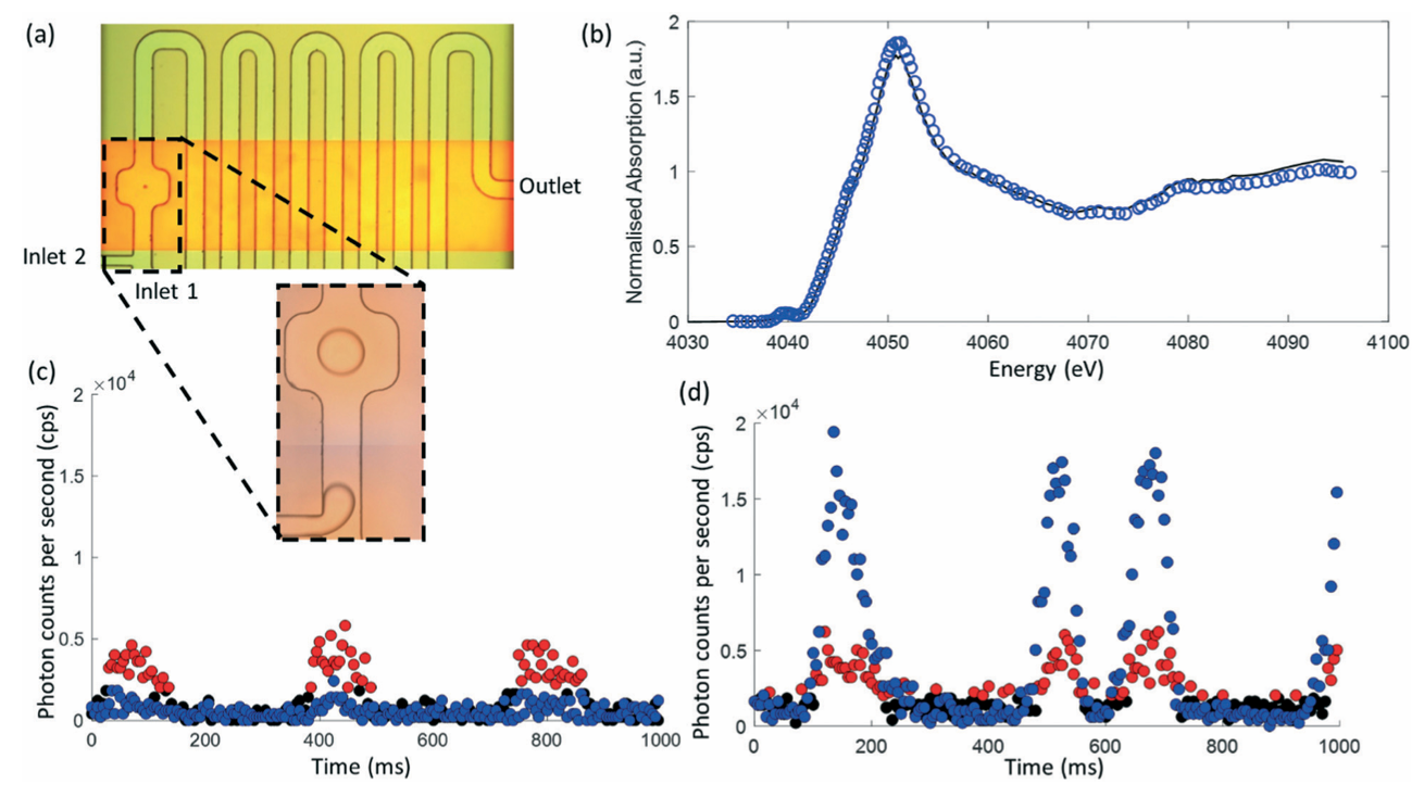
Diffusion coefficients and reaction progress measured directly from fluorescence maps and point spectra
Droplet-based operation, with spectra extracted from individual droplets passing through the beam
Demonstrated
Complexation of calcium with EDTA followed at the calcium K-edge on the PHOENIX beamline of the Swiss Light Source, including diffusion and reaction mapping, pH-dependent coordination, and time-resolved spectra from droplets.
Status
Developed with the Paul Scherrer Institute (Swiss Light Source). Devices can be adapted to new channel geometries, reactions and beamline end stations.
We welcome enquiries from beamline scientists and research groups planning in situ experiments on liquids. See Work with us or contact tuncay.alan@monash.edu.
Key publications
Brenker, J.; Henzler, K.; Borca, C. N.; Huthwelker, T.; Alan, T., X-ray compatible microfluidics for in situ studies of chemical state, transport and reaction of light elements in an aqueous environment using synchrotron radiation. Lab on a Chip 2022, 22 (6), 1214–1230.